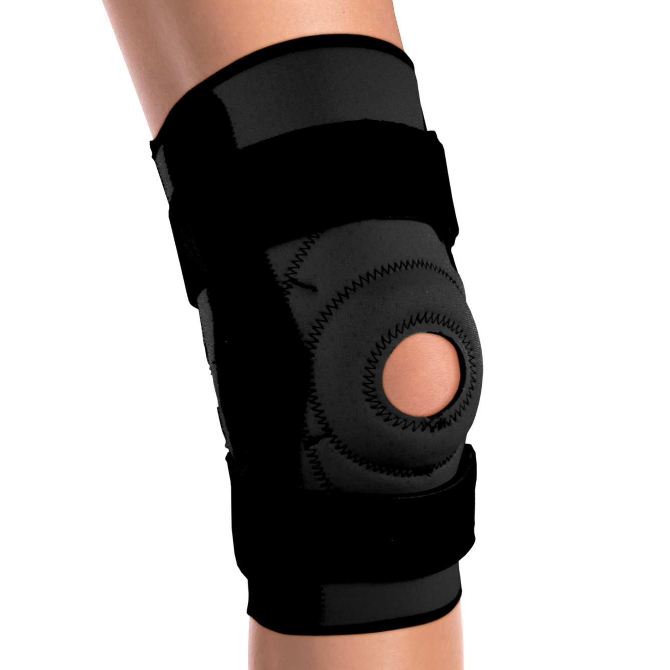
2554 / KNEE BRACE - HINGED BARS
The OTC 2554 knee brace is a heavy duty, controlled stretch support that provides protection and added stability for post-surgical recovery, or for individuals with a history of joint instability. The rigid hinged metal bars provide excellent side-to-side support, yet allow for free flexion and extension. The elastic sleeve supports the entire knee area and helps reduce painful swelling. This is an excellent product for everyday activities like mild exercise and walking.
Indications Listed Below
• Heavy-duty, breathable elastic fabric provides firm, comfortable support
• Lightweight rigid metal hinged bars provide excellent medial-lateral control and allow for free flexion and extension
• Thigh and calf encircling straps are reversible and adjustable to help brace maintain its proper position throughout the day
Product Features
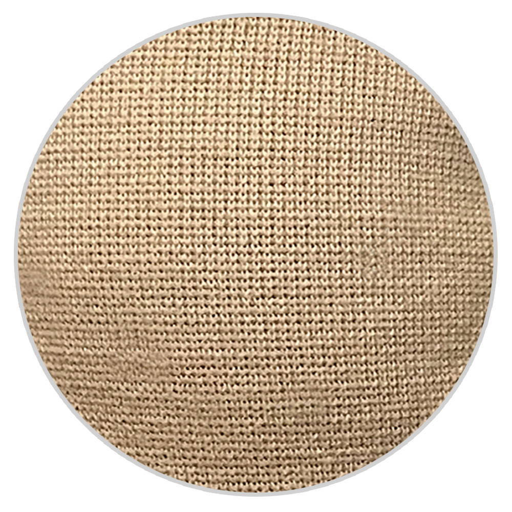
Circular Knit
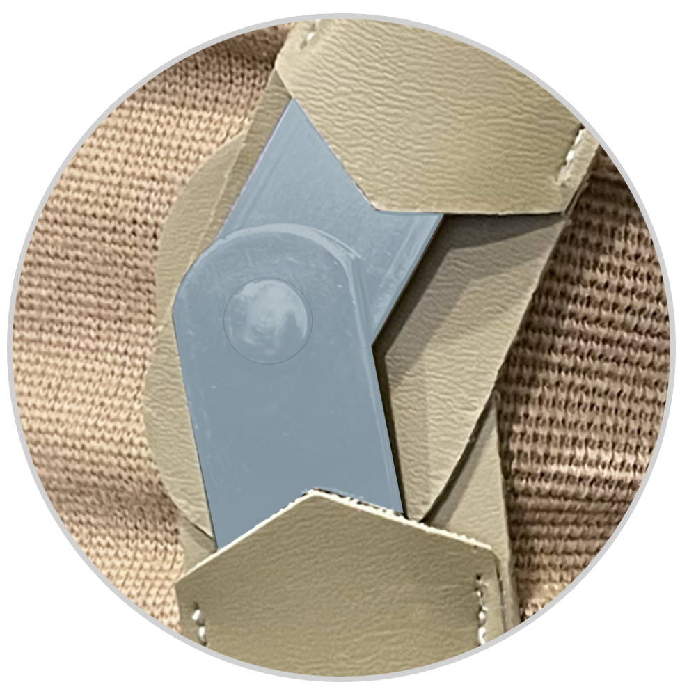
Hinged Bars
Lightweight rigid metal hinged bars provide excellent medial-lateral control and allow for free flexion and extension
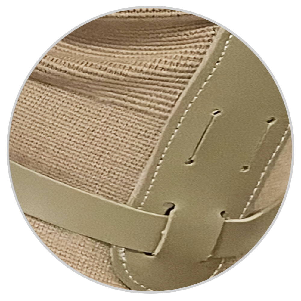
Encircling straps
How to Measure for and Apply Knee Brace
| SIZE | MEASURE AROUND THE BEND OF THE KNEE |
|---|---|
| X-SMALL | 9.5"- 11.5" (24.1 - 29.2 CM) |
| SMALL | 11.5"- 13" (29.2 - 33 CM) |
| MEDIUM | 13"- 14.75" (33 - 37.5 CM) |
| LARGE | 14.75"- 16.5" (37.5 - 41.9 CM) |
| X-LARGE | 16.5"- 18.57" (41.9 - 47 CM) |
| 2X-LARGE | 18.5"- 21" (47 - 53.3 CM) |
| 3X-LARGE | 21"- 23.5" (53.3 - 59.7 CM) |
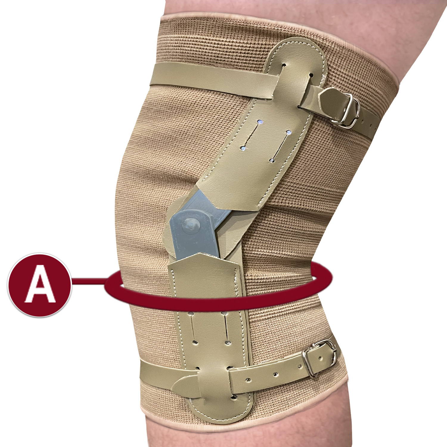
Measuring Instructions
A. Measure around the bend of the knee
Application Instructions
Brace is set for wear on the right knee. If worn on the left, reverse the order of the encircling straps.
1. Remove the hinged bars from their casings and shape them to the medial and lateral contours of the knee.
2. Reinsert the bars into the casings. The longer hinged bar should be positioned at top, and the round rivet head facing the outside.
3. Slip the brace up over the knee until the pivot of the hinge is in line with the bend of the knee. Fasten encircling straps snug.
4. When properly applied, the brace should fit snug but not so tight that it deeply depresses the skin.
Medical Applications
Review the accompanying chart to determine the product that best suits your needs. On the left, you will find a variety of injuries that OTC products are specifically designed to treat and prevent. On the top, you will find the product numbers of all OTC Knee braces, supports, and stabilizers. If a red box is present where the column and row intersect, your injury is treated by the associated product.
| 0141 | 0142 | 0143 | 0144 | 0306 | 0308 | 0309 | 0310 | 0311 | 0312 | 0314 | 0315 | 0326 | 9910 | 9916 | 2415 | 2416 | 2422 | 2422MG | 2425 | 2435 | 2436 | 2540 | 2541 | 2542 | 2553 | 2555 | 2543 | 2544 | 2545 | 2546 | 2548 | 2549 | 2550 | 2554 | |
|---|---|---|---|---|---|---|---|---|---|---|---|---|---|---|---|---|---|---|---|---|---|---|---|---|---|---|---|---|---|---|---|---|---|---|---|
| Arthritis | |||||||||||||||||||||||||||||||||||
| Bursitis | |||||||||||||||||||||||||||||||||||
| Compression & Support | |||||||||||||||||||||||||||||||||||
| Collateral Ligament Support | |||||||||||||||||||||||||||||||||||
| Cruciate Ligament Support | |||||||||||||||||||||||||||||||||||
| Joint Weakness | |||||||||||||||||||||||||||||||||||
| Hamstring Strain or Injury | |||||||||||||||||||||||||||||||||||
| Heat Reduction | |||||||||||||||||||||||||||||||||||
| Inflammation | |||||||||||||||||||||||||||||||||||
| Lateral Pressure Syndrome | |||||||||||||||||||||||||||||||||||
| Lateral Patella Syndrome | |||||||||||||||||||||||||||||||||||
| Medial/Lateral Cartilage Support | |||||||||||||||||||||||||||||||||||
| Medial/Lateral Instability | |||||||||||||||||||||||||||||||||||
| Medial Collateral Ligament Rupture | |||||||||||||||||||||||||||||||||||
| Meniscus Injury | |||||||||||||||||||||||||||||||||||
| Osgood Schlatter Syndrome | |||||||||||||||||||||||||||||||||||
| Osteoarthritis | |||||||||||||||||||||||||||||||||||
| Patellar Misalignment/Instability | |||||||||||||||||||||||||||||||||||
| Patellar Tendon Sprains and Strains | |||||||||||||||||||||||||||||||||||
| Patellar Tendonitis | |||||||||||||||||||||||||||||||||||
| Patellar Fracture or Dislocation | |||||||||||||||||||||||||||||||||||
| Patellar Tendon Rupture | |||||||||||||||||||||||||||||||||||
| Post-Operative Rehabilitation | |||||||||||||||||||||||||||||||||||
| Post-Trauma Discomfort | |||||||||||||||||||||||||||||||||||
| Quadriceps Tendonitis | |||||||||||||||||||||||||||||||||||
| Quadriceps Rupture | |||||||||||||||||||||||||||||||||||
| Runner's Knee/Chondromalachia | |||||||||||||||||||||||||||||||||||
| Sprains and Strains | |||||||||||||||||||||||||||||||||||
| Thigh Pain | |||||||||||||||||||||||||||||||||||
| Traumatic Knee Injuries |
CRUCIATE AND COLLATERAL LIGAMENT INJURIES
The conditions shown below may not be treated by the product listed on this page. Please view the above Medical Applications Chart to determine what conditions this page's associated product treats.
ACL Injuries
The anterior cruciate ligament (ACL) helps maintain knee stability by preventing the tibia (shin bone) from sliding forward beneath the femur (thigh bone). It can be injured in any number of ways, for example: changing direction rapidly, slowing down while running, landing from a jump, and direct injury (such as in a football tackle).

SYMPTOMS
• A “popping”sound noted when injured
• Knee swelling within 6 hours
• Joint instability
• Pain on the medial (inner) side of the knee
PCL Injuries
Posterior cruciate ligament (PCL) injuries disrupt knee joint stability because the tibia can sag backwards. The PCL is usually injured by hyperextension (overextending the knee), or a direct blow to the flexed knee (the position of the knee when you bend the leg).

SYMPTOMS
• Knee swelling and tenderness in the space behind the knee (popliteal fossa)
• Joint instability
• Joint pain
MCL Injuries
The medial collateral ligament (MCL) is located at the inner side the knee joint. The MCL connects the femur to the tibia and provides stability to the inner side of the knee. Injuries to the MCL are usually caused by contact on the inside of the knee.

SYMPTOMS
• Sharp pain on the medial side (inside) of the knee
LCL Injuries
The lateral collateral ligament (LCL) is located at the outer side of the knee joint. The LCL connects the femur to the lateral bone in the lower leg, the fibula, and stabilizes the outer side. Injuries to the LCL are usually caused by contact to the outside of the knee.

SYMPTOMS
• Pain and tenderness along the outside of the kneecap (patella)
• Possible swelling
• Chronic pain and weakness
KNEE ANATOMY

soft tissues of the knee
A. Quadriceps Muscles
The large muscle group found in front of the thigh that traverses the femur and terminates at the supra-patellar tendon. The quadriceps muscles allow the knee to extend or straighten out.
B. Supra-Patellar Tendon
Attaches to the quadriceps muscles to the patella (kneecap).
C. Menisci (Medial and Lateral Meniscus)
Fibrous cartilage pads that distribute weight and provide a smooth surface for the joint to move on.
D. Infra-Patellar Tendon
Attaches the tibia to the patella.

Ligaments of the knee
A. PCL (Posterior Cruciate Ligament)
Attaches at the back of the tibia and the front of the femur. Prevents dislocation of the femur in a forward direction.
B. MCL (Medial Collateral Ligament)
Connects the femur to the tibia and provides stability to the inner side of the knee.
C. ACL (Anterior Cruciate Ligament)
Attaches at the back of the femur and the front of the tibia. Limits rotation and forward movement of the tibia.
D. LCL (Lateral Collateral Ligament)
Connects the femur to the fibula and stabilizes the outer side of the knee.